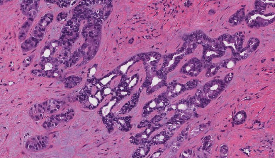
How Cholangiocarcinoma Develops in the Liver
Cholangiocarcinoma typically arises from the epithelial cells lining the bile ducts. While it is classified as a rare cancer, its prevalence has been increasing in certain populations. Cholangiocarcinoma is categorized based on the location of the tumor within the bile duct system:
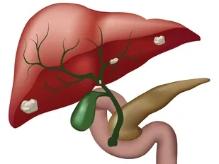
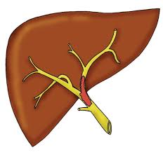
- Intrahepatic Cholangiocarcinoma (ICC) – This type develops in the bile ducts within the liver. Its growth can remain unnoticed for a long time because of the absence of symptoms in its early stages.
- Extrahepatic Cholangiocarcinoma (ECC) – This cancer develops in the bile ducts outside the liver but still within the bile duct system, often leading to bile duct obstruction.
- Perihilar Cholangiocarcinoma (Klatskin Tumors) – This subtype occurs at the junction where the right and left hepatic bile ducts meet before entering the small intestine. These tumors cause significant bile duct blockage and are considered more challenging to treat.
Mechanisms of Development:
- Chronic Inflammation of the Bile Ducts: Long-term inflammation of the bile ducts is one of the primary factors leading to cholangiocarcinoma. This inflammation, often caused by autoimmune diseases or chronic infections, leads to a microenvironment conducive to cancer formation. Chronic conditions such as Primary Sclerosing Cholangitis (PSC) and Biliary Atresia significantly raise the risk of developing cholangiocarcinoma. In PSC, ongoing inflammation causes scarring and narrowing of the bile ducts, which increases the chances of cancer formation over time.
- Cirrhosis of the Liver: Cirrhosis, often caused by chronic alcohol use or chronic viral infections like Hepatitis B or C, leads to extensive liver damage. The constant liver regeneration in cirrhosis increases the potential for errors in cellular reproduction, which can, in turn, result in the development of cholangiocarcinoma.
- Genetic Mutations: Cholangiocarcinoma can also develop due to mutations in certain genes that regulate cell growth and death. These mutations can result from both inherited and acquired genetic changes, which enable abnormal cell proliferation within the bile ducts. For example, mutations in the KRAS, IDH1, BRAF, and TP53 genes have been observed in a large number of cases, particularly in intrahepatic cholangiocarcinoma. These mutations can be targets for new therapeutic strategies, including targeted therapies and immunotherapy.
- Chronic Parasitic Infections: In certain regions, parasitic infections like Clonorchis sinensis and Opisthorchis viverrini are known to cause chronic inflammation in the bile ducts, significantly raising the risk of cholangiocarcinoma. These parasites can introduce harmful substances that promote cellular mutations and bile duct damage over time.
Risk Factors and Population at Higher Risk
Several factors can increase the risk of developing cholangiocarcinoma, especially concerning its association with the liver. These include:
- Age and Gender: Cholangiocarcinoma is more commonly diagnosed in individuals over 50, with men being more frequently affected than women.
- Chronic Liver Diseases: Conditions such as cirrhosis, PSC, chronic hepatitis, and chronic alcoholic liver disease are well-established risk factors.
- Family History: A family history of liver cancers or other types of cancers can increase an individual’s risk of developing cholangiocarcinoma. Genetic predisposition plays a role, although this is relatively rare.
- Previous Gallstones or Gallbladder Disease: Individuals with a history of gallstones or gallbladder disease may have an increased likelihood of developing cholangiocarcinoma.
- Bile Duct Cysts and Genetic Disorders: Certain genetic conditions, such as Caroli’s disease, where bile duct cysts form in the liver, increase the risk of cancer development.
How Cholangiocarcinoma Affects Liver Function
As the tumor grows, it obstructs the bile ducts, which can disrupt bile flow from the liver to the intestines. The liver is responsible for producing bile, which aids in fat digestion, and its blockage leads to a backup of bile in the liver. The result is often jaundice (yellowing of the skin and eyes) and can lead to more severe liver complications, such as liver failure. In advanced stages, the tumor may spread to nearby organs, including the lymph nodes, and metastasize to distant parts of the body.
Symptoms of Cholangiocarcinoma
The signs and symptoms of cholangiocarcinoma are often subtle and can be confused with other conditions, making early diagnosis difficult. However, the following are common symptoms:
- Jaundice: Yellowing of the skin and eyes due to bile buildup in the bloodstream.
- Itchy Skin (Pruritus): Bile salts that accumulate in the skin due to bile duct obstruction cause intense itching.
- Abdominal Pain: Especially in the upper right quadrant, pain can occur as the tumor obstructs bile ducts or presses on surrounding organs.
- Fatigue and Weight Loss: Unexplained weight loss and fatigue are common in many cancers, including cholangiocarcinoma.
- Dark Urine and Pale Stools: The obstruction of bile flow can cause dark-colored urine due to increased bilirubin in the blood, and the stools may become pale or clay-colored due to the lack of bile.
- Fever and Nausea: These can occur due to an infection in the bile ducts (cholangitis) or as a result of liver dysfunction.
Diagnostic Approaches
Diagnosing cholangiocarcinoma requires a multi-faceted approach to confirm its presence and determine its stage. Some of the most common diagnostic methods include:
- Imaging Tests:
- CT Scan: Used to detect tumors, obstruction, and metastasis.
- MRI and MRCP (Magnetic Resonance Cholangiopancreatography): Detailed imaging of bile ducts and liver structures.
- Ultrasound: Non-invasive imaging to identify the presence of abnormal bile duct structures or masses.
- Biopsy: A tissue sample taken from the suspected tumor site allows for histopathological examination to confirm the cancerous nature of the lesion.
- Endoscopic Retrograde Cholangiopancreatography (ERCP): This procedure combines endoscopy and fluoroscopy to visualize the bile ducts and obtain tissue samples for biopsy.
- Blood Tests: Elevated levels of CA 19-9 and liver enzymes can indicate the presence of cholangiocarcinoma, although these markers are not always definitive.
Treatment Options for Cholangiocarcinoma
Cholangiocarcinoma treatment is highly dependent on the stage of the disease, the location of the tumor, and the overall health of the patient. The primary treatment options include:
- Surgical Resection: If the tumor is localized and has not spread beyond the bile duct system, surgery to remove the tumor or even part of the liver may be possible. However, due to the tumor’s location, complete resection can be challenging.
- Liver Transplantation: In cases where the tumor is confined to the liver and surgery is not possible, a liver transplant may be an option. Transplantation can help remove the tumor and address any underlying cirrhosis.
- Chemotherapy and Radiation Therapy: These treatments are often used when the tumor is advanced or surgery is not possible. Chemotherapy can shrink tumors and alleviate symptoms, while radiation therapy targets the tumor to prevent further growth.
- Targeted Therapy: Certain genetic mutations associated with cholangiocarcinoma, such as IDH1 and BRAF mutations, may be treated with targeted therapies designed to block cancer cell proliferation.
- Palliative Care: For advanced stages, where curative treatment is not feasible, palliative care focuses on alleviating symptoms, improving quality of life, and providing support for the patient.
Cholangiocarcinoma remains one of the most challenging cancers to treat, largely due to its tendency to present at an advanced stage. Early diagnosis is critical for improving survival rates, but the lack of early-stage symptoms makes detection difficult. Understanding the risk factors, symptoms, and treatment options is essential for both patients and healthcare providers. Despite its complexity, research into genetic mutations and targeted therapies holds promise for improving outcomes for patients diagnosed with this aggressive liver cancer.