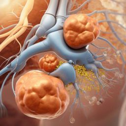
Why Early Diagnosis Matters
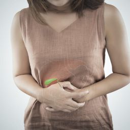
Early detection of gallbladder cancer is critical because the disease is typically asymptomatic in its early stages. By the time symptoms such as abdominal pain, jaundice (yellowing of the skin and eyes), nausea, and weight loss appear, the cancer has often already spread beyond the gallbladder, reducing the chances of successful treatment. In fact, fewer than 20% of people with gallbladder cancer are diagnosed at an early stage, when the disease is still confined to the gallbladder.
When diagnosed early, treatment options such as surgery to remove the gallbladder or tumors can be more effective, leading to better survival rates. On the other hand, if the cancer is detected at a later stage, the options for treatment are limited, and the prognosis is generally poor.
The survival rate for gallbladder cancer patients is closely linked to the stage at diagnosis. Early-stage gallbladder cancer, when confined to the gallbladder, has a much better prognosis compared to advanced stages where the cancer has spread to other organs.
Key Tests for Early Diagnosis of Gallbladder Cancer
While there is no single test that can definitively diagnose gallbladder cancer, a combination of imaging tests, blood tests, and biopsies are necessary for accurate detection. Early detection is vital to prevent the spread of cancer and to increase treatment success. Below are the essential tests that doctors typically recommend for diagnosing gallbladder cancer:
1. Ultrasound

Ultrasound is often the first imaging test performed when gallbladder cancer is suspected. It uses sound waves to create an image of the gallbladder and surrounding structures. While ultrasound is excellent for detecting abnormalities such as gallstones, cysts, or thickening of the gallbladder wall, it is not always conclusive in identifying cancer.
However, an ultrasound can indicate the presence of suspicious masses or tumors, prompting further testing to confirm whether they are cancerous.
- Procedure: The patient lies on an exam table, and a gel is applied to the abdomen. A small handheld device called a transducer is moved across the area to produce images of the gallbladder.
- Benefits: It is non-invasive, widely available, and relatively inexpensive.
- Limitations: Although ultrasound is helpful for identifying gallbladder abnormalities, it may not detect small tumors or cancer that has spread to nearby organs.
2. CT Scan (Computed Tomography)
A CT scan is a more advanced imaging technique that provides detailed cross-sectional images of the body. A CT scan is often used to assess the extent of gallbladder cancer and determine if the cancer has spread to nearby organs, lymph nodes, or other parts of the body.
- Procedure: The patient lies on a table, and a series of X-ray images are taken from various angles. The images are then combined by a computer to create detailed cross-sectional views of the body.
- Benefits: It provides more detailed images than ultrasound and is particularly helpful for detecting tumors that may not be visible on an ultrasound.
- Limitations: CT scans involve exposure to radiation, although the risks are generally considered low.
3. MRI (Magnetic Resonance Imaging)

An MRI scan uses powerful magnets and radio waves to create detailed images of internal structures without the use of radiation. MRI is particularly useful in assessing the liver, bile ducts, and surrounding organs, helping to determine if cancer has spread.
- Procedure: The patient lies on a table inside a large, tube-like machine. The machine uses magnetic fields and radio waves to create detailed images of the body.
- Benefits: MRI provides high-resolution images, especially of soft tissues, which makes it effective for detecting tumors in the gallbladder or surrounding areas.
- Limitations: MRI machines can be claustrophobic for some patients, and the procedure may be more expensive than other imaging tests.
4. Endoscopic Ultrasound (EUS)
Endoscopic ultrasound (EUS) is a specialized type of ultrasound performed through the endoscope, a flexible tube that is inserted through the mouth or rectum. EUS is used to obtain detailed images of the gallbladder, bile ducts, and surrounding tissues.
- Procedure: A thin, flexible tube is inserted through the mouth or rectum, and a small ultrasound probe is used to capture detailed images of the gallbladder and nearby structures.
- Benefits: EUS provides high-resolution images and can help detect smaller tumors or abnormalities that might not be visible with traditional ultrasound.
- Limitations: The procedure is invasive and requires sedation, making it less comfortable for some patients.
5. Blood Tests
Blood tests are commonly used to help diagnose gallbladder cancer and monitor liver function. Certain markers may indicate the presence of cancer or other abnormalities in the gallbladder or liver.
- Liver Function Tests: These tests measure the levels of liver enzymes and other substances in the blood to assess how well the liver is functioning. Abnormal results may suggest liver involvement by cancer.
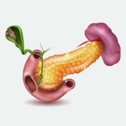
- CA 19-9 Test: This blood test measures the levels of the carbohydrate antigen 19-9 (CA 19-9), a tumor marker that may be elevated in patients with gallbladder cancer or other gastrointestinal cancers. However, high levels of CA 19-9 are not specific to gallbladder cancer and can be elevated in other conditions like pancreatitis or bile duct obstruction.
- CEA Test: The carcinoembryonic antigen (CEA) test is another tumor marker that may be elevated in certain cancers, including gallbladder cancer. Like CA 19-9, CEA is not specific to gallbladder cancer but may indicate the presence of cancer in the body.
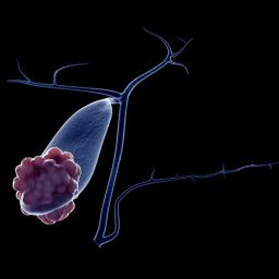
6. Biopsy
A biopsy involves taking a small sample of tissue from the gallbladder or surrounding area to be examined under a microscope for cancer cells. A biopsy is often performed if imaging tests suggest the presence of a tumor.
- Procedure: A biopsy can be done using a needle that is inserted through the skin (percutaneous biopsy) or through the endoscope (endoscopic biopsy) to remove tissue samples.
- Benefits: A biopsy provides the definitive diagnosis of cancer and helps determine the type and stage of the cancer.
- Limitations: Biopsy procedures carry a small risk of infection or injury to surrounding organs, but they are generally safe.
Early diagnosis of gallbladder cancer is critical for improving the chances of successful treatment and increasing survival rates. Since gallbladder cancer often shows no early symptoms, it is essential to rely on a combination of diagnostic tests, including ultrasound, CT scans, MRIs, endoscopic ultrasounds, blood tests, and biopsies, to detect the disease at an early stage.
If you or someone you know is at high risk for gallbladder cancer, it is important to discuss screening options with a healthcare provider. Early detection not only increases the chance of successful treatment but also improves the quality of life during and after treatment. Through regular check-ups and staying vigilant about symptoms, patients can significantly improve their chances of surviving gallbladder cancer.