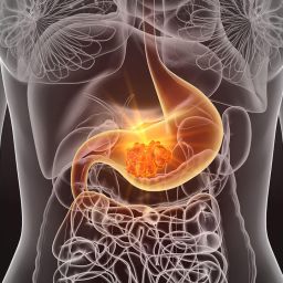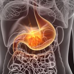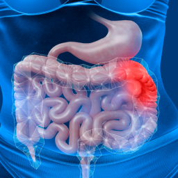
1. Defining Benign and Malignant GISTs
Before exploring the differences, it’s important to first define the terms benign and malignant in the context of GISTs:
- Benign GISTs are non-cancerous tumors. While they can grow and cause symptoms, they typically do not spread to other parts of the body. Benign GISTs generally have a more favorable prognosis and can often be managed with surgical resection alone.
- Malignant GISTs, on the other hand, are cancerous tumors that have the potential to invade surrounding tissues and metastasize (spread) to distant organs. Malignant GISTs are often more aggressive, requiring a combination of surgery, targeted therapies, and close follow-up.
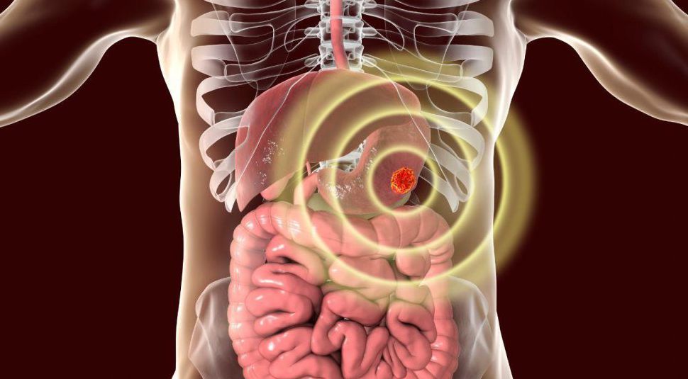
Despite being relatively rare, GISTs are the most common mesenchymal tumor of the GI tract, and the malignant forms can be particularly challenging to manage. The differentiation between benign and malignant GISTs is crucial for deciding on the appropriate course of treatment and predicting the patient’s long-term prognosis.
2. Tumor Characteristics and Risk Factors
a. Tumor Size
One of the most significant factors used to distinguish benign GISTs from malignant ones is tumor size. In general, larger GISTs have a higher likelihood of being malignant. However, size alone is not always a definitive predictor.
- Benign GISTs are typically smaller, often measuring less than 5 cm in diameter.
- Malignant GISTs, especially those that are metastatic, are generally larger than 5 cm and can reach sizes of 10 cm or more, depending on their location and stage.
Tumors larger than 5 cm are more likely to have high mitotic activity (rapid cell division), which is often associated with malignancy.
b. Mitotic Rate and Cellular Proliferation
The mitotic rate, or the rate of cell division, is a key feature used to differentiate benign from malignant GISTs. Tumors with higher mitotic rates are considered to be more aggressive.
- Benign GISTs have a lower mitotic rate, typically under 5 mitoses per 50 high-power fields (HPF).
- Malignant GISTs exhibit a higher mitotic rate, often exceeding 5 mitoses per 50 HPF. This higher rate of cell division leads to the tumor’s growth and potential spread to other organs.
Histopathological analysis is required to measure the mitotic rate. Malignant GISTs are often characterized by rapid cellular turnover and abnormal tissue structures.
c. Tumor Necrosis
Tumor necrosis, or areas of dead tissue within the tumor, is another factor used to classify GISTs.
- Benign GISTs rarely have significant necrosis, and when they do, it is usually less extensive.
- Malignant GISTs often exhibit areas of necrosis, indicating aggressive tumor behavior. Necrosis can be an indication of poor blood supply to the tumor, which is common in rapidly growing or metastasizing tumors.
Necrosis is typically evaluated by pathologists during histological examination and is a hallmark of malignant GISTs.
3. Molecular and Genetic Differences
a. C-kit Mutations
One of the key molecular markers used to distinguish between benign and malignant GISTs is c-kit mutation status. The c-kit gene encodes for a protein that plays a role in cell growth and survival.
- Benign GISTs are often associated with mutations in the c-kit gene, particularly in exon 11, which typically lead to a less aggressive behavior. These tumors are often responsive to tyrosine kinase inhibitors (such as imatinib), which target the c-kit mutation.
- Malignant GISTs, particularly those that are high-risk or metastatic, can also harbor c-kit mutations, but the mutation type and the gene’s response to treatment can vary. Some malignant GISTs may be resistant to imatinib, especially if there are secondary mutations in c-kit or other mutations in related genes.
b. PDGFRA Mutations
Another key mutation found in GISTs is the PDGFRA (platelet-derived growth factor receptor alpha) gene. While most GISTs harbor c-kit mutations, a subset of tumors has PDGFRA mutations, particularly in exon 18.
- Benign GISTs with PDGFRA mutations often have a more indolent course and a lower risk of metastasis.
- Malignant GISTs with PDGFRA mutations can be more aggressive, although these mutations tend to be less common than c-kit mutations. The prognosis and treatment response in these cases depend on the mutation’s exact nature and whether the tumor exhibits resistance to imatinib.
Both c-kit and PDGFRA mutations are identified through molecular testing and gene sequencing.
c. Other Genetic Markers
Some GISTs are associated with other genetic mutations, such as SDH (succinate dehydrogenase) gene mutations, which are more common in familial cases and are associated with a different biological behavior.
- Benign GISTs with SDH mutations can exhibit slow growth and low metastatic potential.
- Malignant GISTs with SDH mutations, particularly in children and younger adults, may demonstrate a more aggressive behavior.
4. Histopathological Examination and Risk Stratification
Histopathology plays a vital role in determining whether a GIST is benign or malignant. Pathologists examine tissue samples for various characteristics, including tumor size, mitotic rate, necrosis, and genetic markers.
GISTs are typically categorized into three risk groups based on their histopathological features:
- Low-risk GISTs: These tumors are smaller in size (less than 5 cm) and have a low mitotic rate (less than 5 mitoses per 50 HPF). They are less likely to recur or metastasize.
- Intermediate-risk GISTs: These tumors may have moderate mitotic rates and size, making them more likely to recur or metastasize, but they still have a relatively good prognosis with treatment.
- High-risk GISTs: These tumors are larger, have a high mitotic rate, show significant necrosis, and may already have spread to distant sites (metastasized). They require more aggressive treatment and closer follow-up.
Risk stratification based on histopathology helps clinicians determine the most appropriate treatment approach and tailor follow-up protocols.
5. Clinical Behavior and Symptoms
a. Benign GISTs
Benign GISTs are often asymptomatic and may be discovered incidentally during routine exams or imaging studies for unrelated conditions. However, as the tumor grows, it can cause symptoms such as:
- Abdominal pain or discomfort
- Nausea and vomiting
- Gastrointestinal bleeding (though rare)
Because benign GISTs grow slowly and are less likely to spread, surgical removal of the tumor is often sufficient for treatment. Once resected, benign GISTs typically do not recur.
b. Malignant GISTs
Malignant GISTs, in contrast, tend to be more aggressive and present with a range of symptoms, often due to metastasis or local tumor invasion:
- Abdominal mass: May be palpable or visible on imaging.
- Weight loss and fatigue: Common in advanced stages.
- Gastrointestinal bleeding: Can occur due to the tumor’s location and vascularity.
- Signs of metastasis: Symptoms related to the spread of the tumor, such as pain in the liver or lungs, may also be present.
Malignant GISTs require a multidisciplinary approach to treatment, including surgery, targeted therapy, and close monitoring for recurrence or metastasis.
6. Treatment Strategies for Benign and Malignant GISTs
The treatment approach for GISTs varies significantly depending on whether the tumor is benign or malignant.
a. Treatment of Benign GISTs
For benign GISTs, surgery is the mainstay of treatment. If the tumor is small and has clear, negative surgical margins, the likelihood of recurrence is low. In some cases, adjuvant therapy with imatinib (a tyrosine kinase inhibitor) may be used, especially if the tumor has c-kit mutations.
b. Treatment of Malignant GISTs
Malignant GISTs typically require more aggressive treatment strategies, including:
- Surgical resection: The primary approach for localized malignant GISTs is surgical removal. However, achieving clear margins can be challenging if the tumor is large or has invaded nearby organs.
- Targeted therapy: For high-risk or metastatic GISTs, targeted therapy with imatinib or other tyrosine kinase inhibitors is essential. These drugs target the c-kit and PDGFRA mutations that drive tumor growth.
- Radiation therapy: Radiation is typically not effective for GISTs but may be used in specific cases of local recurrence or to alleviate symptoms.
- Chemotherapy: Conventional chemotherapy is generally not effective for GISTs, and it is reserved for cases with secondary cancers or when resistance to targeted therapies occurs.