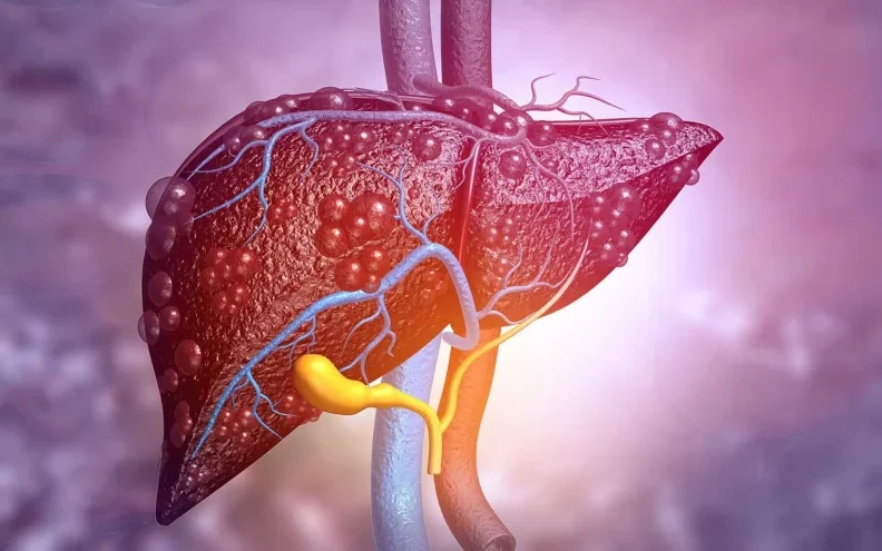
Symptoms of Liver Hemangioma
In many instances, liver hemangiomas do not cause any noticeable symptoms. These benign tumors are often discovered accidentally during imaging exams for other health concerns. However, when symptoms do present, they are often related to the size of the tumor, its location in the liver, or complications such as rupture or bleeding.
1. Abdominal Pain
The most common symptom associated with liver hemangiomas is abdominal pain, particularly in the upper right side of the abdomen where the liver is located. The pain is typically dull, persistent, and can be exacerbated by eating large meals or pressure on surrounding organs. The severity of the pain can vary depending on the size of the hemangioma and whether it is pressing against other structures in the abdomen.
2. Nausea and Vomiting
Some individuals with larger liver hemangiomas may experience nausea and vomiting. This symptom is more likely when the tumor interferes with the digestive process or causes a sensation of fullness due to the pressure it exerts on surrounding organs. These symptoms may also occur if there is bleeding within the hemangioma.
3. Bloating and Early Satiety
As the hemangioma grows, it can occupy space in the abdomen and cause a sensation of bloating or early satiety. People with larger hemangiomas may feel full after eating only small amounts of food, which can affect their appetite and eating habits.
4. Jaundice
In rare cases, liver hemangiomas can cause jaundice, which is characterized by a yellowing of the skin and eyes. Jaundice occurs when the hemangioma obstructs the bile ducts, preventing bile from flowing out of the liver properly. This can result in the buildup of bilirubin in the bloodstream, which leads to the yellowish tint in the skin and eyes.
5. Bleeding or Rupture
One of the most serious complications associated with liver hemangiomas is bleeding or rupture, although it is relatively rare. If the hemangioma grows large or becomes fragile, it may rupture, leading to significant internal bleeding. This is a medical emergency that requires immediate attention, as it can lead to shock or other life-threatening complications.
Causes and Risk Factors of Liver Hemangiomas
The exact cause of liver hemangiomas is not fully understood. However, there are several potential factors that may contribute to the formation and growth of these benign tumors.
1. Hormonal Factors
There is evidence suggesting that hormones, particularly estrogen, may play a role in the development and growth of liver hemangiomas. These tumors are more common in women, especially those who are pregnant or taking hormonal medications such as oral contraceptives or hormone replacement therapy (HRT). During pregnancy, the elevated levels of estrogen may encourage the growth of existing hemangiomas or even lead to the development of new ones.
2. Genetic Factors
Though liver hemangiomas typically occur sporadically, certain genetic factors may contribute to their development. For example, people with von Hippel-Lindau disease, a rare genetic disorder that increases the risk of tumors in various organs, may have a higher chance of developing hemangiomas in the liver. Family history may also play a role, although genetic factors alone are not sufficient to explain the development of these tumors.
3. Vascular Malformations
Liver hemangiomas are essentially abnormal blood vessel formations, which means that any condition or developmental abnormality that affects blood vessel formation during fetal development can potentially lead to the development of liver hemangiomas. These vascular malformations are typically present from birth, though they may not be discovered until later in life.
4. Age and Gender
Liver hemangiomas are more commonly diagnosed in middle-aged women, particularly those between the ages of 30 and 50. This age and gender predisposition is thought to be related to the influence of estrogen on blood vessel growth. The condition is much rarer in men and children, with most cases becoming apparent in adulthood.
5. Other Risk Factors
While most liver hemangiomas are sporadic and idiopathic (without clear cause), certain environmental and lifestyle factors may indirectly increase the likelihood of developing these tumors. For example, heavy alcohol use, liver disease, or long-term exposure to toxic substances could, in theory, increase the risk of developing abnormal liver growths, though no direct link has been definitively established.
Diagnosis of Liver Hemangiomas
Liver hemangiomas are most often diagnosed through imaging techniques. Since these tumors typically do not cause symptoms, they are commonly discovered incidentally when a person undergoes an ultrasound, CT scan, or MRI for another health issue.
1. Ultrasound
Ultrasound is usually the first imaging test used to detect liver hemangiomas. It is a non-invasive and relatively inexpensive method that uses sound waves to create an image of the liver. While it can effectively detect liver hemangiomas, ultrasound may not always provide enough detail to differentiate between hemangiomas and other liver conditions.
2. CT Scan (Computed Tomography)
A CT scan provides more detailed imaging and is commonly used to assess the size, shape, and structure of a liver hemangioma. A contrast-enhanced CT scan, where a special dye is injected into the bloodstream, can offer more clarity and detail, helping doctors distinguish hemangiomas from other types of liver lesions.
3. MRI (Magnetic Resonance Imaging)
MRI is considered the gold standard for diagnosing liver hemangiomas. It provides high-resolution images that clearly reveal the characteristics of the hemangioma. An MRI is especially useful in differentiating liver hemangiomas from other potential liver tumors, such as hepatic cysts or malignancies. It can also help doctors evaluate the size and location of the hemangioma and plan appropriate treatment.
4. Biopsy
In rare cases, a biopsy may be performed to confirm the diagnosis of a liver hemangioma. However, biopsies are typically not required unless there is uncertainty about the nature of the tumor. Liver hemangiomas are generally benign, so invasive procedures like biopsies are not commonly used unless necessary for ruling out other conditions.
Treatment of Liver Hemangiomas
For most individuals, liver hemangiomas do not require treatment. If the hemangioma is small, asymptomatic, and does not cause complications, a watch-and-wait approach with regular monitoring is usually sufficient. However, in some cases, intervention may be necessary, especially if the hemangioma grows large or causes symptoms.
1. Observation and Monitoring
In many cases, especially when the liver hemangioma is small and asymptomatic, the best course of action is simply to monitor the tumor over time. Regular follow-up with imaging tests, such as ultrasounds or MRIs, helps doctors track the tumor’s growth. If the hemangioma remains stable and does not cause any issues, no further treatment may be required.
2. Hormonal Treatment
Since hormonal factors such as estrogen may contribute to the growth of liver hemangiomas, adjusting or discontinuing hormone-based medications may be recommended in certain cases. For instance, if a woman is using oral contraceptives or hormone replacement therapy (HRT), her doctor may advise stopping these medications to prevent further growth of the hemangioma.
3. Surgical Intervention
Surgical removal of the hemangioma is typically considered only when the tumor becomes symptomatic, large, or causes complications such as bleeding or pain. Surgery may involve partial liver resection, where the portion of the liver containing the hemangioma is removed. This procedure is generally considered safe, though it is reserved for cases where the benefits outweigh the risks.
4. Embolization
In certain cases, particularly when surgery is not feasible or when the tumor is inaccessible, embolization may be used. This procedure involves blocking the blood vessels supplying the hemangioma, causing it to shrink over time. Embolization is typically performed through a catheter inserted into a blood vessel in the groin.
5. Radiation Therapy
Radiation therapy may be considered in rare cases where the hemangioma is large, symptomatic, or cannot be treated by other means. Radiation therapy uses high-energy radiation to shrink or destroy the abnormal blood vessels in the hemangioma. This is generally not a first-line treatment, but it may be an option for individuals who are not candidates for surgery.
6. Liver Transplantation
In extremely rare instances, when a liver hemangioma leads to liver failure or severe complications that cannot be managed through other treatments, liver transplantation may be considered. This option is typically reserved for patients with other underlying liver diseases in addition to the hemangioma.
Liver hemangiomas are the most common type of benign liver tumors, often discovered incidentally during imaging for unrelated conditions. These tumors are generally asymptomatic and do not require treatment. However, in rare cases, when they become symptomatic or cause complications, medical intervention may be necessary. Treatment options range from simple observation to more invasive procedures such as surgery or embolization.
Understanding the potential causes, symptoms, and treatment methods for liver hemangiomas is essential for individuals diagnosed with the condition. Regular monitoring, lifestyle modifications, and timely intervention when required can help manage this condition effectively and prevent complications.